I. What are microbes?Cellular
| FUNGI | |
 |
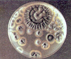 |
| Psilocybe cyanescens, a potent species widespread through western Europe and prolific in the Pacific Northwest of North America. | Penicillium colonies |
 |
|
| Psilocybe stuntzii, a 'magic' mushroom and Galerina autumnalis, a deadly poisonous mushroom growing in wood chips. Note that they are growing so close together that they are actually touching! | |
| BACTERIA | |
 click for enlargement click for enlargement |
 |
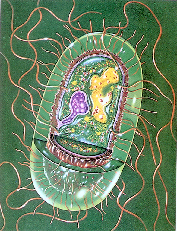
| The ultrastructure of Salmonella, a bacterium thta causes human diseases such as typhoid fever and food poisoning. | |
 |
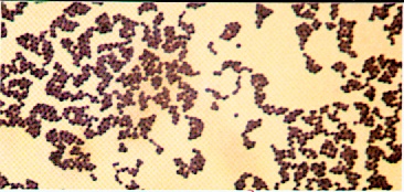 |
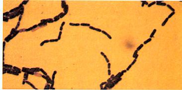
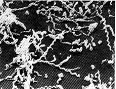
| Bacillus megaterium, a rod-shaped bacterium in chains. Gram stain (x600). | Staphylococcus aureus. Note the gram-positive spheres in irregular clusters. Gram stain (x1,000). |
 |
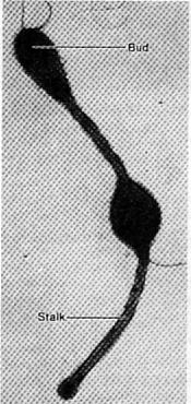 |
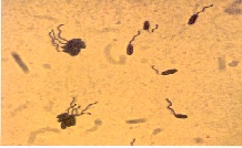
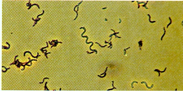
| Rhodospirillum rubrum. Phase contrast (x500). | Hyphomicrobium with stalk and bud, electron micrograph with negative staining. |
 |
 |
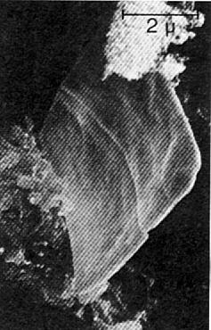
| Walsby's square bacterium. | Spiroplasma, SEM (x13,000). |
 |
|
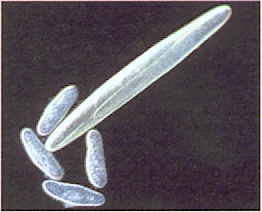
| Epulopiscium fishelsoni. This photograph, taken with pseudo dark field illumination, shows E.fishelsoni at the top of the figure dwarfing paramecia at the bottom (x200). | |
| VIRUS | |
 |
 |
| Ebola | Hepatitis B virus |
 |
|
| Herpesvirus | |
| PROTISTS | 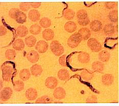 |
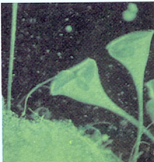 |
| The protozoan Trypanosoma brucei gambiense, blodd smear (x1,000), one cause of African sleeping sickness. | Stentor. The ciliated protozoa are extended and actively feeding, dark-field microscopy (x100). | 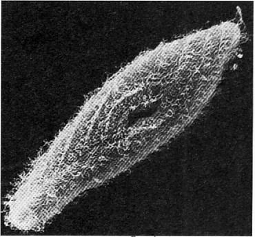 |
| Coordination of Ciliary Activity. A scanning electron micrograph of Paramecium showing cilia (x1,500). The ciliary beat is coordinated and moves in waves across the protozoan's surface, as can be seen in the photograph. | |